Basic Info
high-end materials science research, semiconductor material analysis and precision metal product inspection, providing high-performance imaging quality and rich expansion functions. It adopts infinity corrected plan apochromatic objective lens groups, featuring extremely high resolution and color restoration, allowing clear observation of nano-scale metal microstructures, adapting to cutting-edge material research and high-end quality inspection needs. It supports multi-mode imaging including transmitted light, reflected light and polarized light imaging, meeting diversified sample analysis requirements.
Customer reviews

Robert Williams · Metallurgical Consultant
After 15 years in metallurgical consulting, I can confidently say this is the most versatile microscope I've used. The brightfield/darkfield switching mechanism allows instant transition between observation modes - crucial when examining complex multi-phase materials. The 5-axis mechanical stage provides precise control for documenting specific microstructural features. For clients who need documented evidence, the camera port produces publication-quality micrographs without additional processing.
Sarah Johnson · Research Lab Technician
Working in a university metallurgy lab, we needed equipment that could withstand heavy student use while maintaining precision. This microscope has proven incredibly durable - the fine focus mechanism still operates smoothly after two years of daily use by multiple researchers. The interchangeable objectives allow quick adaptation between different sample types, and the built-in scale reticle has been invaluable for our quantitative analysis projects.
Michael Chen · Forensic Engineer
For fracture analysis in forensic investigations, this microscope provides the perfect balance of portability and performance. The 20x to 2000x magnification range covers all our needs, from initial fracture surface examination to detailed inclusion analysis. The anti-reflection coatings on lenses significantly reduce glare from polished metal samples. We've been able to identify fatigue striations and crack propagation directions with much greater confidence since upgrading to this system.
David Reynolds · Quality Control Manager
In our aerospace component manufacturing facility, this microscope has revolutionized our failure analysis process. The polarized light capability reveals stress patterns in metal alloys that our previous equipment couldn't detect. We've significantly reduced false negatives in our inspections since implementing this system. The ergonomic design also reduces operator fatigue during long inspection sessions - a major improvement over traditional models.
Emily Carter · Materials Scientist
As a materials scientist, I've used several microscopes over the years, but this high-resolution research-grade metallurgical microscope stands out. The image clarity at 1000x magnification is exceptional, allowing me to analyze grain boundaries and phase distributions with unprecedented detail. The LED illumination provides consistent brightness without overheating samples. What impressed me most was the seamless integration with our digital imaging system - the color accuracy in captured images matches what I see through the eyepieces.
Product Description
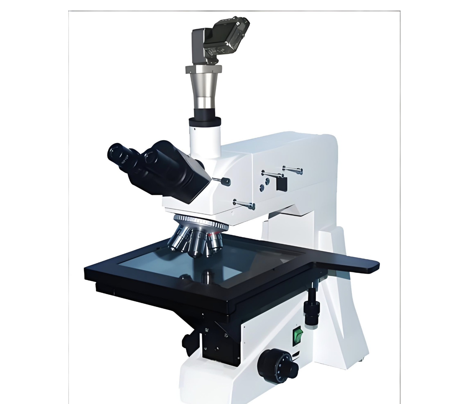
Product features:
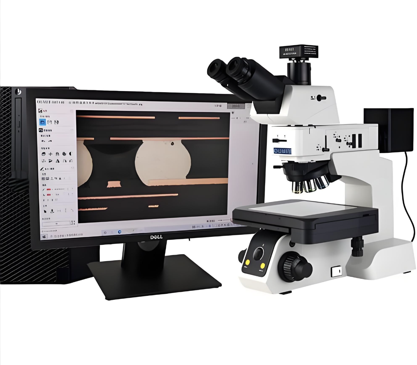
The core strengths of this product lie in its ultra-high resolution and multi-mode imaging capabilities. It adopts an infinity corrected optical system, effectively reducing spherical aberration and chromatic aberration of traditional objective lenses, achieving an ultra-high imaging resolution of 0.1μm, and clearly observing nano-scale metal grains and semiconductor wafer defects. It is equipped with a trinocular tilting observation head, supporting simultaneous observation by two people and computer imaging acquisition. It is equipped with a 12V 100W halogen transmitted light source and fiber optic epi-illumination reflected light source, allowing flexible switching between transmitted and reflected imaging modes to adapt to transparent and opaque metallographic samples. The supporting Image-Pro Premier software supports advanced image analysis, 3D reconstruction, grain statistics and quantitative analysis, meeting scientific research-level data processing needs. The body adopts a shock-proof optical platform design, helping to minimize the impact of external vibration on imaging quality, and comes with a 3-year full machine warranty and free on-site installation and debugging services.
Product specifications:
Detailed technical parameters include a magnification range of 50x-2000x, with 50x/100x/200x/500x infinity corrected plan apochromatic objective lens groups, paired with 10x/16x high-point eyepieces. The trinocular tilting observation head has a pupil distance adjustment range of 55-75mm and a 0-360° rotation function. The mechanical stage measures 240×200mm, with X-Y axis coarse and fine adjustment knobs, supporting a maximum sample thickness of 50mm. The imaging resolution reaches 0.1μm, adopting a dual light source system: 12V 100W halogen transmitted light and fiber optic epi-illumination reflected light. The overall dimensions are 580×450×650mm, with a net weight of 28kg, supporting 110/220V wide-range AC power supply. It is equipped with a USB 3.0 interface for high-speed data transmission, compatible with mainstream scientific research-grade imaging cameras, and comes with a 3-year full machine warranty and 1-year free consumable replacement service.
Product application:
It applies to cutting-edge metal material research in college materials science research institutes, wafer defect detection in semiconductor chip manufacturing, microstructure analysis of high-temperature alloys in the aerospace field, micro-topography detection of cutting edges in precision tool manufacturing, and pole piece structure analysis of new energy vehicle batteries. Specific scenarios include researching the grain growth mechanism of new high-entropy alloys by scientific research teams, detecting metal wiring defects on wafer surfaces in semiconductor factories, analyzing the high-temperature oxide layer structure of turbine blades in aviation enterprises, and detecting micro-wear of cutting edges in precision tool factories, supporting researchers and high-end manufacturing enterprises to complete high-precision microstructure analysis and quality control.
Frequently Asked Questions (FAQ)
Q:What are the key features of a high-resolution research-grade metallurgical microscope?
A:Key features include: 1) Ultra-high resolution (up to 0.1μm) for microstructure analysis, 2) Advanced LED/epi-illumination for opaque samples, 3) Motorized XYZ stage with 1μm precision, 4) Compatibility with EBSD and EDX detectors.
Q:How does a research-grade metallurgical microscope differ from industrial models?
A:Research-grade models offer: 1) Higher NA objectives (0.9+) for superior resolution, 2) Advanced image analysis software, 3) Vibration-resistant platforms, 4) Polarization/DIC capabilities for material characterization, whereas industrial models prioritize durability over precision.
Q:What magnification range is typical for high-end metallurgical microscopes?
A:Standard ranges: 1) 50x-1000x for routine analysis, 2) 2000x with oil immersion for nanoscale features, 3) Optional 5000x with specialized optics. Most systems include 5x-100x plan-apochromatic objectives.
Q:Why choose a motorized metallurgical microscope for materials research?
A:Motorization provides: 1) Automated z-axis stacking for 3D reconstruction, 2) Precise repeatable measurements (±0.5μm), 3) Remote operation for cleanroom/harsh environments, 4) Integration with AI-based particle analysis systems.
Q:What sample preparation is required for metallurgical microscopy?
A:Essential steps: 1) Mounting in conductive resin, 2) Precision polishing (0.05μm finish), 3) Etching with HNO3 or other reagents, 4) Carbon coating for non-conductive samples. Preparation quality directly impacts imaging clarity.


 Post RFQ


 Chat
Chat


 ALL CATEGORIES