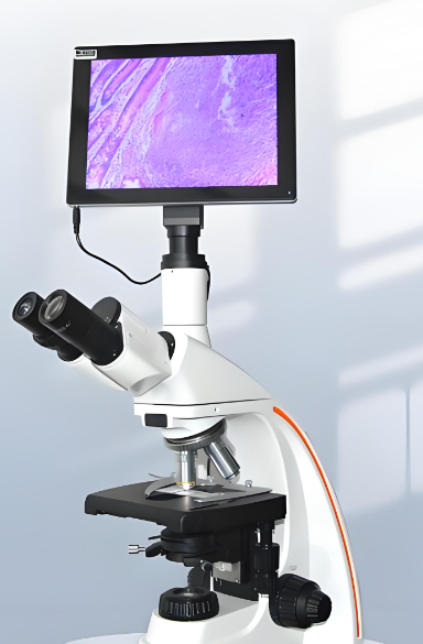
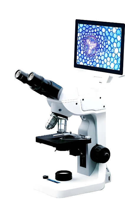


The core advantage lies in professional biomedical adaptability and high-precision imaging. It adopts confocal stereoscopic scanning technology, which can clearly observe the three-dimensional structure of biological samples, and supports fluorescence imaging mode, which can be used for observation and scanning of fluorescently labeled cells and tissue sections. It is equipped with electric fine-tuning focusing system and motorized XY sample stage, which can realize automatic scanning and stitching of a wide range of samples, improving scientific research efficiency. It is compatible with fluorescence LED lighting and white light lighting, which can meet the observation needs of different types of biological samples. It is equipped with professional biomedical image analysis software, which can realize cell counting, structure measurement, image stitching and other functions. The fuselage is made of die-cast aluminum and medical-grade ABS materials, which meet the hygiene standards of biological laboratories and are easy to clean and disinfect.
The adjustable magnification range is 40x-1600x, adopting confocal stereoscopic 3D scanning imaging scheme, with native resolution of 7680*4320 8K UHD, supporting real-time high-definition imaging and fluorescence imaging. The working distance ranges from 15mm to 100mm, adapting to conventional biological sample slides and petri dishes. The stroke of the motorized XY sample stage is 100mm*80mm, which can realize automatic sample scanning and panoramic imaging. The fuselage is made of die-cast aluminum and medical-grade ABS materials, with a total weight of about 12.3kg and dimensions of 580mm (length) * 450mm (width) * 320mm (height). It is powered by AC 100-240V mains power, supporting electric focusing and electric sample stage control, compatible with professional biomedical image analysis software, and equipped with USB 3.2, HDMI 2.1 and professional camera interfaces, which can connect to computers and scientific research equipment.
It is mainly applicable to pathological section analysis, cell three-dimensional structure research, biological specimen observation, medical teaching demonstration and other scenarios. It can provide professional 3D biological microscopic observation tools for medical colleges, scientific research institutes and hospital pathology departments, helping researchers obtain complete three-dimensional structure information of biological samples, improving the accuracy and efficiency of medical research and pathological diagnosis.


 Post RFQ


 Chat
Chat


 ALL CATEGORIES