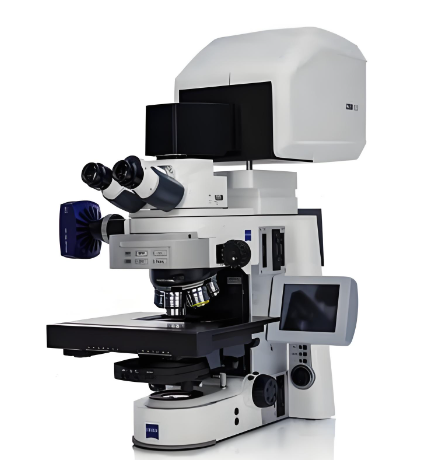
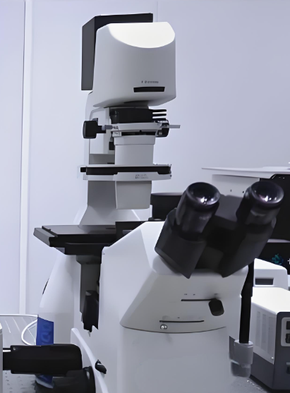


The core advantages of this confocal laser microscope lie in its ultra-high resolution and three-dimensional imaging capability, which can clearly capture the internal structure of samples with a Z-axis resolution of 0.1μm. It adopts a stepless adjustable confocal pinhole design, which can flexibly adjust the imaging depth and field of view according to testing needs. The cooled sCMOS sensor has high sensitivity and low noise, enabling clear imaging of weak fluorescent signals. The professional analysis software supports 3D structure reconstruction, fluorescence intensity quantification and cell movement trajectory tracking, effectively solving the pain points of traditional microscopes that cannot perform deep internal structure imaging.
The main specifications include: overall dimensions of 1100mm (length) × 750mm (width) × 600mm (height), net weight of 120kg, maximum scanning speed of 30fps, and the maximum imaging field of view is 1024×1024 pixels. It supports four standard laser wavelengths, covering common fluorescent marker excitation requirements. The objective lens set includes 10x, 20x, 40x and 100x oil immersion lenses, with a numerical aperture of up to 1.49. The supporting analysis software can realize offline data analysis and report generation, supporting export of multiple file formats including TIFF, JPEG and CSV.
This product is mainly suitable for cutting-edge scientific research scenarios such as 3D tissue structure reconstruction, cell membrane dynamics observation, material surface topography analysis and nano-structure detection. It is widely used in universities, national key laboratories, biomedical research institutions and material science research centers. For example, neurobiologists can use it to observe the synaptic connection structure of brain tissues in three dimensions, providing important experimental data for the study of brain neural circuits.


 Post RFQ


 Chat
Chat


 ALL CATEGORIES